

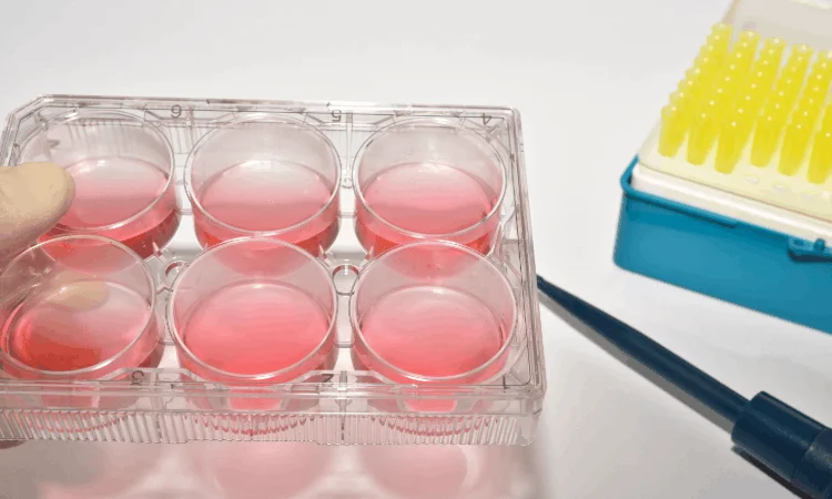
Cytotoxicity Testing: Demonstrating the Safety of Medical Devices at the Cellular Level
The first and most critical step in the biocompatibility assessment of medical devices is determining whether the device has a toxic effect on cells. The cytotoxicity test determines whether a medical device or material is safe at the cellular level by evaluating its effects on cell viability, proliferation capacity, and cellular morphology. This test is conducted in accordance with the ISO 10993-5 standard and constitutes one of the cornerstones of biocompatibility assessment. Our laboratory conducts cytotoxicity tests in vitro using methods such as MTT, NRU, and XTT, as well as direct contact and agar overlay techniques, in accordance with GLP and GMP principles.
The Scientific Basis and Importance of the Cytotoxicity Test
Cytotoxicity refers to the totality of toxic effects that a substance or material exerts on cells. These effects may manifest as cell death (necrosis or apoptosis), cell membrane damage, decreased metabolic activity, arrest of cell proliferation, and morphological changes. The cytotoxicity test enables the quantitative or qualitative measurement of these effects in vitro. The test’s most significant advantage is that it does not require the use of animals and provides rapid, cost-effective, and reproducible results.
The cytotoxicity test is considered the first step in biocompatibility assessment. If a device is cytotoxic, further testing (sensitization, irritation, implantation) is generally unnecessary; because cytotoxicity is evidence that the device exhibits serious incompatibility at the cellular level. Therefore, the cytotoxicity test is a critical screening test for both patient safety and the efficient use of the manufacturer’s resources.
Cytotoxicity test results are directly used in the preparation of the device’s biocompatibility dossier, in regulatory submissions (EU MDR 2017/745, FDA 510(k), ISO 10993-1), and in the safety assessment of the device prior to clinical use. Furthermore, the evaluation of the cytotoxic potential of raw materials, additives, mold release agents, and sterilization residues used in the device’s manufacturing process is considered an indicator of production quality.
Scope and Test Methods of the ISO 10993-5 Standard
ISO 10993-5 is the internationally recognized fundamental standard for the in vitro cytotoxicity evaluation of medical devices. This standard provides for the evaluation of cytotoxic effects by applying extracts obtained from the device or the device material itself directly to cell cultures. Three basic test approaches are defined within the scope of the standard: the extraction method, the direct contact method, and the agar overlay (agar diffusion) method.
Extraction Method involves applying extracts prepared from the device in accordance with ISO 10993-12 to cell cultures. This method is ideal for evaluating the cytotoxic potential of the device’s soluble components. Extracts are prepared using polar (serum-containing medium) and non-polar (vegetable oil) solvents; thus, both water-soluble and lipophilic components are evaluated. Extraction temperatures and durations are determined to reflect the device’s clinical usage scenario.
The direct contact method involves placing the device material directly onto the cell culture. This method is particularly suitable for solid devices and surface materials, as it directly evaluates the effects resulting from the device’s physical contact with the cells. The test material is placed on top of the cell monolayer, and cell viability and morphology are assessed after a specified incubation period. The direct contact method is valuable for evaluating the device’s surface properties and local effects in the contact area.
The agar overlay (agar diffusion) method is performed by pouring a layer of agar onto a cell culture and placing the device material on top of this layer. This method allows soluble substances released from the device to diffuse through the agar layer and reach the cells. Agar overlay is particularly suitable for devices with irregular surfaces or those containing particles; it also prevents mechanical damage that could result from direct physical contact between the device and the cells.
Cell Viability Measurement Methods: MTT, NRU, and XTT
Various colorimetric methods are used to measure cell viability in cytotoxicity assays. These methods indirectly determine the number of live cells by measuring cellular metabolic activity or membrane integrity. The three most commonly used methods are MTT, NRU, and XTT.
The MTT assay is based on the principle that MTT tetrazolium salt (3-(4,5-dimethylthiazol-2-yl)-2,5-diphenyltetrazolium bromide) is reduced by mitochondrial dehydrogenase enzymes in cells to form purple-colored formazan crystals. The formazan crystals produced in live cells are dissolved in a suitable solvent (DMSO or isopropanol) and measured spectrophotometrically. The absorbance value is directly proportional to the number of live cells. The MTT assay is a fast, economical, and widely used method.
The NRU (Neutral Red Uptake) assay is based on the ability of live cells to accumulate the neutral red dye in their lysosomes. Live cells actively take up neutral red and store it in their lysosomes; dead cells, however, cannot take up this dye. The dye is extracted with an acidic alcohol solution and measured spectrophotometrically. The NRU assay is used specifically to assess membrane integrity and lysosomal function; it may provide more stable results compared to the MTT assay.
The XTT assay is based on the principle of the reduction of XTT (2,3-bis-(2-methoxy-4-nitro-5-sulfophenyl)-2H-tetrazolium-5-carboxanilide), a tetrazolium salt similar to that used in the MTT assay, by cells. The most significant advantage of XTT is that it forms a water-soluble formazan product; therefore, the dissolution step required in the MTT assay is not necessary. The XTT assay is faster and requires fewer steps; it also exhibits lower cytotoxicity compared to the MTT assay.
The table below summarizes the test methods and cell viability measurement techniques used under ISO 10993-5.
| Test Method | Procedure | Suitable Device Types | Evaluation |
|---|---|---|---|
| Extraction | Application of extracts obtained from the device to cells | All device types (solid, liquid, gel, powder) | Cell viability measurement using MTT, NRU, or XTT |
| Direct Contact | Placement of the device material directly onto the cell monolayer | Solid devices, surface materials, films | Morphological assessment and cell viability measurement |
| Agar Overlay | Placement of the device material on an agar layer | Devices with irregular surfaces, materials containing particles | Diffusion zone and morphological assessment |
| MTT | Measurement of mitochondrial dehydrogenase activity | All cell culture assays | Spectrophotometric measurement of formazan crystals |
| NRU | Measurement of lysosomal neutral red uptake | All cell culture assays | Spectrophotometric measurement of neutral red dye |
| XTT | Tetrazolium reduction (water-soluble formazan) | All cell culture assays | Spectrophotometric measurement of the formazan solution |
Evaluation and Classification of Cytotoxicity Results
Cytotoxicity test results are evaluated according to the criteria defined in ISO 10993-5. The percentage of cell viability is calculated by comparison with the negative control. The generally accepted classification is as follows: If cell viability is above 70 percent, the material is considered “non-cytotoxic.” If cell viability is between 50 and 70 percent, the material is classified as “mildly cytotoxic”; in this case, additional testing or optimization may be required. If cell viability is below 50 percent, the material is classified as “significantly cytotoxic,” and it is concluded that the device is not suitable for clinical use.
Morphological evaluation is an important complement to numerical measurements. The shape, size, cytoplasmic granulation, membrane integrity, and intercellular junctions of the cells are evaluated under a microscope. For example, cell rounding, cytoplasmic retraction, vacuolization, and detachment of the cell layer are morphological indicators of cytotoxicity. These observations are interpreted in conjunction with quantitative data to reach a final decision.
Cytotoxicity test results are a key component of the biocompatibility report to be used in the device’s regulatory submission. If the device is not cytotoxic, testing can proceed to other biocompatibility tests (sensitization, irritation, implantation). If the device is cytotoxic, the manufacturer must review the material composition, production process, or sterilization method. In this case, our laboratory provides scientific consultation to the manufacturer regarding alternative materials, coatings, or decontamination steps.
Conducting Cytotoxicity Tests in Our Laboratory
In our GLP- and GMP-certified laboratory, we conduct cytotoxicity tests in accordance with ISO 10993-5, in full compliance with international standards. Our testing process begins with a comprehensive preliminary evaluation. The device’s material composition, manufacturing process, sterilization method, and clinical usage scenario are examined in detail. Based on this information, the most appropriate test method (extraction, direct contact, or agar overlay) and cell viability measurement technique (MTT, NRU, or XTT) are determined.
The extraction process is performed in accordance with ISO 10993-12. Using specific volumes of polar (serum-containing culture medium) and non-polar (vegetable oil) solvents per unit surface area or mass of the device, extraction is carried out at temperatures and for durations that reflect clinical use. Polar and non-polar extracts are tested separately. Cell cultures are prepared using internationally recognized cell lines (L-929 mouse fibroblasts, BALB/3T3, V79, or human skin fibroblasts). Cells are propagated under appropriate culture medium and incubation conditions (37°C, 5% CO₂).
After the test material or extract is applied to the cells, it is incubated for a specific period (typically 24–72 hours). At the end of incubation, cell viability is measured using the MTT, NRU, or XTT assay. The results are interpreted by comparing them to negative and positive controls. High-density polyethylene (HDPE) or high-purity glass is used as the negative control, while solutions containing organotin (dibutyltin dilaurate) or phenol are used as the positive control. These controls are critical for verifying the sensitivity of the test system.
One of our laboratory’s key advantages is that we offer customized testing strategies for different device types. For liquid-form devices (gels, creams, solutions), direct application is used; for solid devices, direct contact or extraction is used; for materials containing particles, agar overlay is used; and for devices with irregular surfaces, modified extraction protocols are used. This flexibility ensures that the unique characteristics of each device are properly evaluated.
Test results are presented in a comprehensive test report format compliant with ISO 10993-5. The report includes sections on the test method, cell line, extraction conditions, cell viability data, morphological observations, statistical analyses, and interpretation. Our reports meet the requirements of the EU MDR (2017/745), FDA 510(k), ISO 10993-1, and other regulatory frameworks.
Frequently Asked Questions
One of the most common questions we receive regarding cytotoxicity testing is which test method is suitable for which device. The choice depends on the device’s physical form and clinical usage scenario. For liquid and gel-based devices, extraction or direct application may be appropriate; for solid devices, direct contact or extraction; and for materials containing particles, agar overlay may be suitable. Our laboratory evaluates the characteristics of your device to recommend the most appropriate method.
The difference between MTT, NRU, and XTT assays is also frequently asked. MTT measures mitochondrial activity and requires the dissolution of formazan crystals. NRU assesses lysosomal function and provides information on membrane integrity. XTT, on the other hand, is faster and requires fewer steps because it produces a water-soluble formazan product. The choice of method depends on the device’s characteristics and the laboratory’s preference; however, all methods are validated and internationally recognized.
Regarding the test duration, the total time—including cell culture preparation, extraction, incubation, and measurement steps—is generally 2–4 weeks. Including reporting and quality assurance reviews, this period may extend to 4–6 weeks. As for which devices are subject to cytotoxicity testing, the answer is that, in accordance with ISO 10993-1, a cytotoxicity assessment is mandatory for all medical devices that come into contact with the skin, mucous membranes, blood, or tissue. This is the first and most fundamental step in biocompatibility assessment.
Finally, there is often curiosity about what should be done if a device is found to be “cytotoxic” as a result of cytotoxicity testing. In this case, the manufacturer must review the material composition, the manufacturing process (mold release agents, solvents, additives), and the sterilization method. Our laboratory offers advanced analytical methods—such as extraction fractionation, chemical analysis, and alternative material testing—to identify the source of the problem. The goal is to ensure the device’s safety and compliance with regulatory requirements.
A cytotoxicity test provides scientific evidence of your medical device’s safety at the cellular level. A rigorous cytotoxicity study conducted in accordance with ISO 10993-5:2009 forms the foundation of your device’s biocompatibility profile and represents the first critical step in the regulatory approval process. Our laboratory evaluates your device’s cellular compatibility to the highest scientific standards using MTT, NRU, and XTT methods, as well as direct contact and agar overlay techniques. To be your trusted partner on your medical device’s biocompatibility journey, contact us.


